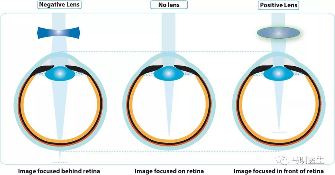
Over the last four decades, numerous animal models have provided valuable insight into the mechanisms underlying emmetropization and refractive error development. Much of the knowledge on vision-dependent changes in ocular growth has emanated from animal experiments in which either the quality of image formed on the retina is degraded (known as form-deprivation [FD]), or the focal point of the image is altered with respect to the retinal plane (known as lens induced defocus). Both FD and lens induced defocus result in abnormal eye growth and development of refractive errors. This section summarizes the attributes of experimental ametropias derived from these two visual manipulations, their differences, and significance for understanding refractive error development in humans.
在过去的40年里,许多动物模型为正视化和屈光不正发展机制提供了有价值的见解。关于依赖视觉引起的眼球生长的许多知识源于动物实验,在动物实验中,视网膜上形成的图像质量下降(称为形觉剥夺[FD]),或者图像的焦点相对于视网膜平面改变(称为镜片诱导离焦)。FD和镜片诱导离焦都会导致眼睛异常生长和屈光不正。本节总结了从这两种视觉操作中得出的实验性屈光不正的属性,它们的区别以及对理解人类屈光不正的重要性。
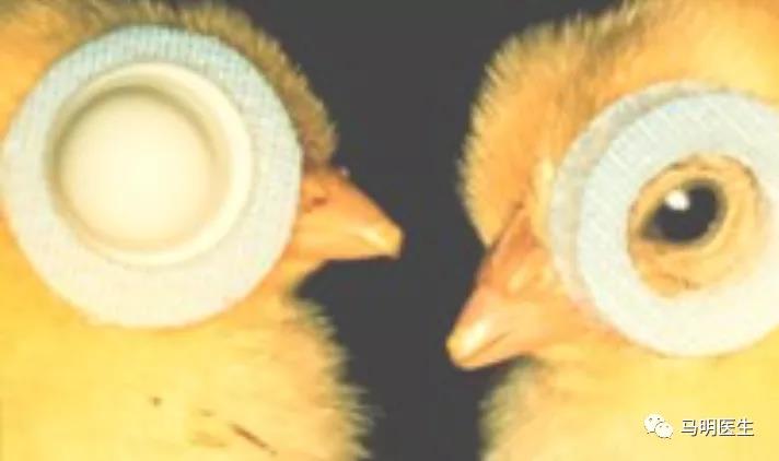
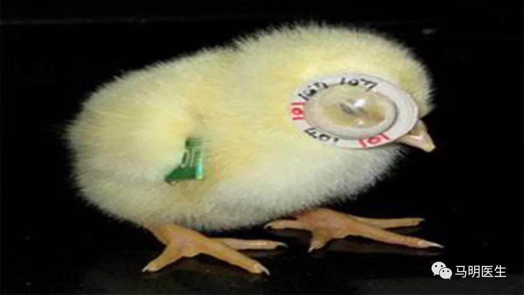
FD is the most commonly used experimental paradigm to model axial myopia in animals. Depriving the retina of form or patterned vision through eyelid suture, or translucent diffusers consistently produces axial myopia (Fig. 4.5). The use of noncontact translucent diffusers offers a more reliable representation of ocular changes with FD since they do not induce corneal changes (unlike eyelid fusion techniques). The ocular changes observed in response to FD clearly illustrate that degrading retinal image quality can produce robust myopic changes. Schaeffel et al. proposed that FD is an open-loop condition, in which myopia develops as a result of uncoordinated ocular growth due to reduced retinal image contrast (or mid-range spatial frequency vision) and the absence of visual feedback related to the effective refractive state of the eye.
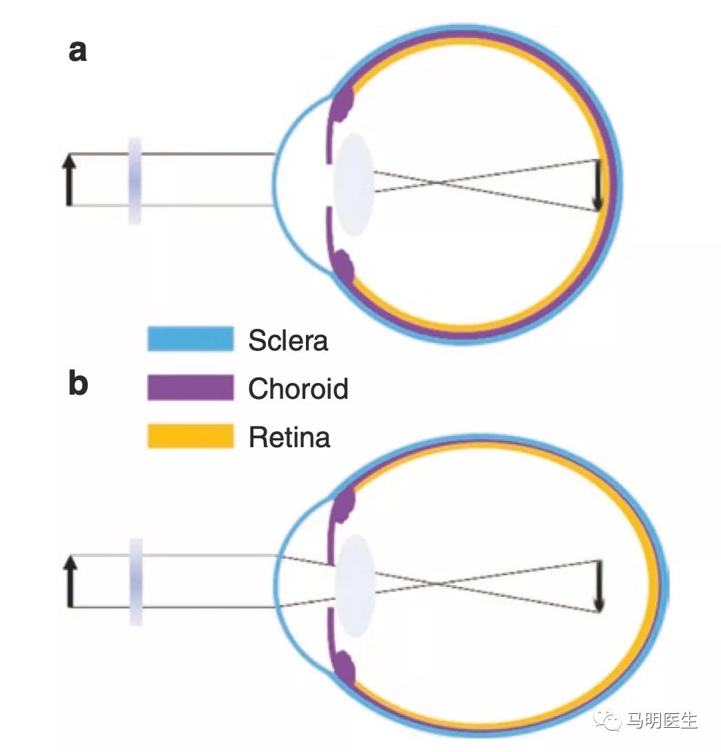
Fig. 4.5 Ocular compensation for form-deprivation (FD). (a) A diffuser causes nondirectional blur and a reduction in contrast of the retinal image. (b) The absence of visual feedback related to the effective refractive state of the eye causes a thinning of the posterior choroid and an increase in ocular growth, resulting in myopia
图4.5形觉剥夺(FD)的视觉补偿。(a)柔光镜会引起非方向性模糊,并降低视网膜图像的对比度。 (b)由于缺乏与眼睛有效屈光状态相关的视觉反馈,导致后脉络膜变薄,眼球生长加快,从而导致近视
FD是目前最常用的动物轴性近视模型。通过缝合眼睑或半透明柔光镜剥夺视网膜的形态或有图案的视觉,持续产生轴性近视(图4.5)。使用非接触半透明柔光镜提供了一个更可靠的显示FD的眼部变化,因为它们不会引起角膜的变化(不像缝合眼睑技术)。在FD中观察到的眼部变化清楚地说明了降低视网膜图像质量可以产生明显的近视变化。Schaeffel等人提出FD是一种开环状态,近视是由于视网膜图像对比度降低(或中等空间频率视觉),以及与眼的有效屈光状态相关的视觉反馈缺失导致眼球生长不协调而形成的。
The myopic response to FD varies among different animals. It is generally greatest in chickens (−9 D after 5 days of FD), followed by tree shrews (−8 D after 12 days in young animals) and guinea pigs (−6.6 D after 11 days), and is less pronounced and much more variable in marmosets (−8 D after 4.5 weeks) and rhesus monkeys (~−5 to −6 D after 17 weeks). Variations observed between individual studies and animal models may be due to differences in experimental paradigms, the duration and extent of FD, inherent ocular ana- tomical variations, and/or differences in susceptibility to environmental myopia. Nevertheless, the myopic response to FD is conserved across a wide range of animal species (including fish, rabbit, mouse, and kestrel). In all species, axial myopia is predominantly caused by a significant elongation of the vitreous chamber, along with thinning of the choroid and the sclera. Few studies have also reported changes in corneal curvature and lens thickness with FD. Interestingly, ocular conditions that cause varying degrees of visual deprivation in humans such as ptosis, congenital cataract, corneal opacity, and vitreous hemorrhage are associated with myopia, which may result from mechanisms similar to FD myopia observed in animals.
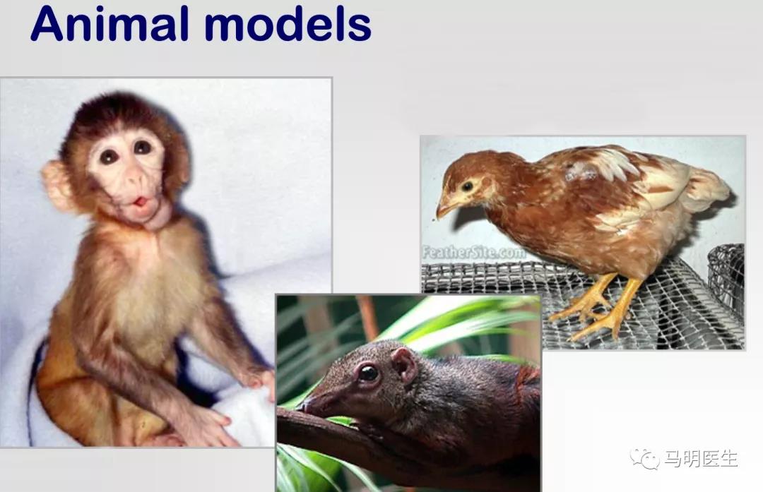
FD对近视的反应在不同的动物之间有所不同。通常小鸡最明显(经过5天的FD产生−9D), 其次是树鼩(幼龄动物12天后−8D)和豚鼠(11天后−6.6D), 不太明显的是狨猴(4.5周后为-8D)和恒河猴(17周后为约-5至-6D)。在个别研究和动物模型之间观察到的差异可能是由于实验模式的差异,FD的持续时间和程度、眼部的固有解剖变化和/或对环境近视的敏感性差异所致。然而,尽管如此,对FD的近视反应在许多物种中都存在(包括鱼类,兔子,小鼠和红隼)。在所有物种中,轴向近视主要是由玻璃体腔的显着延长以及脉络膜和巩膜变薄引起的。少数研究也报道了FD改变角膜曲率和晶状体厚度。有趣的是,引起人类不同程度形觉剥夺的眼部疾病如上睑下垂、先天性白内障、角膜混浊和玻璃体出血都与近视有关,这可能是由于类似于在动物中观察到的FD近视的机制所致。
FD myopia is a graded phenomenon, where increasing degrees of image degradation are positively correlated with the magnitude of induced axial myopia. In addition, the effects of FD declines with age. Younger chicks, macaques, tree shrews, and marmosets show greater ocular changes in response to image degradation compared to older animals, potentially due to age-related reductions in sensory processing of blur stimuli or changes in scleral growth.
FD近视是一种渐变现象,图像质量减低程度的增加与诱发轴性近视的大小正相关。此外,FD的作用随年龄增长而减弱。幼鸡、猕猴、树鼩和绒猴与年龄较大的动物相比,在图像退化时表现出更大的眼部变化,这可能是由于模糊刺激的感官处理或巩膜生长的变化引起的。